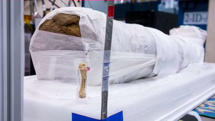
Researchers Used Advanced X-Rays to Reveal Secrets of an Ancient Egyptian Mummy—Without Unwrapping It

Ancient mummies, being so well preserved, can tell us a lot about the lives of those in the past. But studying their secrets usually involves unwrapping the mummies—and possibly damaging them in the process. While researchers have long used CT (computerized tomography) scans to skirt this issue, the X-rays aren’t detailed enough to uncover everything about the artifacts inside.
Fortunately, more comprehensive X-ray technologies are emerging. The U.S. Department of Energy’s Argonne National Laboratory, southwest of Chicago, houses the Advanced Photon Source (APS), a light source facility that produces much more intense X-ray beams than what we’d get for a broken bone. “The difference is akin to the difference between a laser and a light bulb,” APS physicist Jonathan Almer tells Mental Floss.
Since regular X-rays show contrast based on density, they’re useful for things like identifying cracks—which are filled with air—in dense bone. APS X-rays, on the other hand, show contrast based on crystal lattices. Basically, each crystalline material has its own lattice: a repeating molecular pattern that differs in type and size from the lattices of other materials. Because these lattices are so distinct, the information that researchers can glean from APS beams is much more specific than what a standard X-ray would reveal. “For example, we can distinguish high-calcium bone from low-calcium bone due to differences in lattice size, or the amount of carbon in steel,” Almer explains.
Almer and a team of scientists at Northwestern University recently turned the APS’s high-resolution beams on a 1900-year-old mummy excavated in Hawara, Egypt, in 1911. A preliminary CT scan had suggested that the remains belonged to a 5-year-old child—likely female, based on the portrait found with the mummy. Because her skeleton is intact, the researchers think she may have died of disease, rather than bodily harm. The CT scan also helped researchers decide which areas of the mummy to target with the APS beams, cutting the X-ray process from two weeks down to about 24 hours.
Using the new X-rays, the team shed light on a couple key mysteries about the mummy. Tiny wire pins that punctured the cloth turned out to be made from a “modern dual-phase steel,” suggesting that they were added in recent decades to keep the wrappings secure. The other mysterious material is much older (and more surprising): The amulet resting on the skeleton’s abdomen was created from a calcium carbonate mineral called calcite. Northwestern University research professor Stuart Stock, who co-authored the accompanying study in the Journal of the Royal Society Interface, explained in a press release that calcite wasn’t an especially common material for amulets of this ilk. Therefore, researchers may soon be able to track down details about when and where it originated.
Overall, the study is an example of how technological advances in one field can impact research in a completely different one. It’s also evidence that new technologies can further archaeological research without sacrificing precious artifacts.