Scientific Achievement
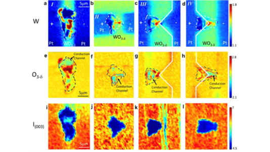
We are the first to image the conduction channel formed in a memristor using an X-ray microscope with both structural and chemical contrast. Our in situ multimodal imaging method allows us to directly see how local electric fields create field-induced defect structures in a model device.
Significance and Impact
Memristors in neuromorphic computers emulate biological synapses, but intrinsic randomness can lead to poor numerical accuracy. By in situ imaging channel formation, we showed that the oxygen vacancy concentration and pathway can be controlled by geometrical means. Our work was highlighted in G. Pacchioni, Nature Review Materials 7, 594 (2022).
Research Details
- Fabricated model memristors from single crystal WO3 films
- Imaged the structure and chemical composition before and after channel formation with the X-ray microscope at Sector 2 of the APS.
Argonne National Laboratory seeks solutions to pressing national problems in science and technology by conducting leading-edge basic and applied research in virtually every scientific discipline. Argonne is managed by UChicago Argonne, LLC for the U.S. Department of Energy’s Office of Science.
The U.S. Department of Energy’s Office of Science is the single largest supporter of basic research in the physical sciences in the United States and is working to address some of the most pressing challenges of our time. For more information, visit https://energy.gov/science.