From nano to macro: upscaling imaging of the mouse brain
Solving this problem is important because it could provide new insights into vertebrate brain structure and connectivity.
Given this importance, scientists have explored numerous techniques, ranging from visible light sources to electron microscopy. Human brain studies have been limited, however, by two major problems: the density of synaptic organizations and the enormous number of connections involved, making mapping of all these connections infeasible. Much research therefore has focused on mouse brains, and significant progress has been made in conducting micrometer-scale studies with electron microscopy. Most of these studies have been done on segmented sections of the brain. X-ray microscopy has also been widely used for neuroanatomy studies of significant portions of mouse brains, including 1µm resolution on whole mouse brains, An open question, however, is whether these studies can be extended to the macroscopic scale.
A team of researchers from Argonne National Laboratory and Northwestern University recently collaborated on tackling this question. Their work is featured as a lead article in the April issue of the Journal of Applied Crystallography.
The first requirement for imaging increasingly thick specimens is to have sufficient image contrast and an acceptable radiation dose.
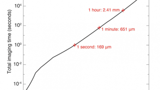
“Knowing the density of the specimen is vital,” said Zichao (Wendy) Di, a computational mathematician with a joint appointment in Argonne’s Mathematics and Computer Science Division and Advanced Photon Source. Once the density is known, the next step is estimating the optimum amount of photon energy. For example, at lower photon energies, the specimen can become too absorptive for optimum imaging; but at higher photon energies, the contrast decreases, requiring a larger number of photons per pixel; see Fig 1.
The researchers used x-ray ptychography, a computational method involving the generation of images by processing many diffraction patterns from different positions of a specimen. It offers the advantage of being “dose efficient,” but it still raises a number of questions. Does the dose of a 3D reconstruction require the same integral dose as a conventional 2D micrograph? How many photons are needed to illuminate the entire object? Can one achieve imaging beyond the depth-of-focus limit?
To answer these questions, the researchers began with a computational model that gives excellent agreement with thin-specimen observations for imaging features at a specific contrast and resolution. They then incorporated corrections for thick-specimen imaging. They also took into account the coherent flux expected at various x-ray energies from the Advanced Photon Source Upgrade at Argonne and the Advanced Light Source Upgrade at Berkeley. With this information the researchers were able to calculate the minimum per-pixel imaging time, as well as the photon energy that minimizes the imaging time, and extrapolate that to whole-specimen 3D imaging times.
The researchers have answered the first part of their original question in the affirmative: Yes, the imaging process can be scaled up. But the second part – can this upscaling be done in a reasonable amount of time – remains challenging.
“Since ptychography involves obtaining diverse patterns from various positions, maximizing the throughput time is critical,” Di said. One possibility that has recently showed promise is staining, which increases the contrast of specific features, without unduly increasing the overall absorption in thick specimens.
Several other issues also need to be addressed, Di noted. “We will need dramatic advances in the available frame rate of detectors with a modest number of pixels. We also will need improved high-speed scanning systems. And we will need ‘smart sampling,’ using artificial intelligence to collect a larger signal where required for critical feature identification and a smaller signal from other regions.”
The path is indeed challenging. But the researchers are optimistic that with new technology and new computational methods, scientists will be able to fully exploit the next generation of diffraction-limited synchrotron light sources.
For further information see the paper by Ming Du, Zichao (Wendy) Di, Doga Gürsoy, R. Patrick Xian, Yevgenia Kozorovitskiy, and Chris Jacobsen, Upscaling X-ray nanoimaging to macroscopic specimens, Journal of Applied Crystallography 54: 386-401, 2021; https://scripts.iucr.org/cgi-bin/paper?S1600576721000194